
IVDD & Spinal Decompression Surgery
Intervertebral Disc Disease (IVDD) can rob your dog of their mobility overnight. Dr. Mark Tonzing utilises advanced, tissue-sparing microsurgical techniques to rapidly decompress the spinal cord, relieve severe pain, and restore your dog's ability to walk.
Preserved Stability
Our preferential use of mini-hemilaminectomies preserves the spinal articular facet joints, maintaining critical structural stability in your dog's spine post-surgery.
Advanced Microsurgery
Accessing the neural canal via targeted pediculectomies and partial corpectomies minimises trauma to surrounding tissues and drastically reduces spinal cord manipulation.
State-of-the-art Safety
Every procedure is supported by on-site 40kW digital X-ray, high-speed pneumatic burrs, and advanced anaesthetic machines with integrated multi-parameter monitors and auto-ventilators for uncompromised safety.
Understanding IVDD & Spinal Compression
Intervertebral Disc Disease (IVDD) occurs when the cushioning discs between the vertebrae of the spine degenerate and rupture (herniate) upward into the spinal canal. Because the spinal canal is a confined, rigid tube of bone, the herniated disc material has nowhere to go but to press violently against the delicate spinal cord.
This compression cuts off nerve signals travelling to the back legs and bladder. The result is sudden, excruciating pain, a wobbly or "drunken" gait (ataxia), dragging of the hind paws, or complete paralysis.
Time is critical. The longer the spinal cord remains compressed, the higher the risk of permanent, irreversible nerve damage. If your dog is losing the ability to stand, this is a surgical emergency.
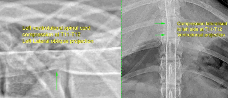
Advanced imaging is required to pinpoint the exact location and severity of the disc herniation prior to surgical decompression.
The BoneVet Surgical Approach: Tissue-Sparing Spinal Decompression
To relieve the pressure on the spinal cord, a window must be drilled through the vertebral bone to extract the ruptured disc material. However, how that window is created makes a massive difference to your dog's postoperative comfort and long-term spinal stability.
The Standard Hemilaminectomy
Traditionally, surgeons perform a standard hemilaminectomy. This involves drilling away a large portion of the side of the vertebra, which almost always necessitates the removal of the articular facet joint (the structural joint connecting two vertebrae together).
While this provides a wide view of the spinal cord for the surgeon, sacrificing the facet joint can lead to localised spinal instability (subluxation), increased risk of complications due to formation of a laminectomy membrane and increased postoperative pain due to extensive tissue disruption. Hemilaminectomy has also been shown to be less effective at removing disc material that has extruded ventrally (underneath the spinal cord), which is a common occurrence in IVDD cases.
Pediculectomy in preference to Hemilaminectomy
At BoneVet, Dr. Tonzing preferentially utilises a pediculectomy. Instead of removing the entire side of the vertebral arch including the articular facets, we use a high-speed pneumatic burr to drill a small, highly precise "keyhole" window slightly lower down below the articular facets—straight through the pedicle of the vertebra.
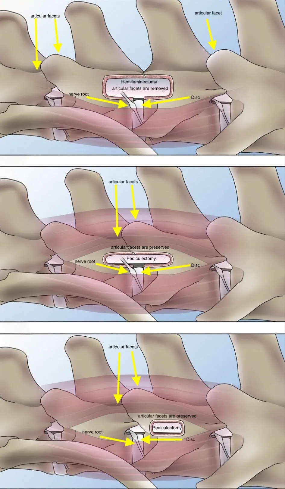
- Preserved Articular Facets: The structural joints of the spine are left completely intact, eliminating the risk of surgically-induced spinal instability.
- Less Tissue Trauma: A smaller access window means less muscle dissection and significantly reduced postoperative pain.
The Role of Partial Corpectomy
In cases where the disc material is wedged underneath the spinal cord (ventral compression), we may perform a partial corpectomy. By drilling a small slot into the vertebral body itself, we gain direct access to the herniated disc material from below. This allows us to extract the disc without having to pull, retract, or manipulate the highly fragile spinal cord—drastically improving neurological recovery outcomes.
The Technical Demand
These tissue-sparing techniques demand a high degree of technical ability. Operating through a keyhole window millimetres away from the spinal cord requires profound spatial awareness, specialised surgical lighting, and exact mastery of high-speed burring techniques. It is a demanding approach, but it yields undeniably superior results for the patient while maintaining spinal stability.
The Neurological Recovery Timeline
Rehabilitating the spine takes patience and strict compliance.
Strict Confinement & Pain Management
Your dog will be discharged once their pain is managed and they are able to urinate (or you are comfortable expressing their bladder). They must be strictly confined to a crate or small pen. The spinal cord is highly inflamed, and absolute rest is required for the swelling to subside. Only assisted sling-walking for toilet breaks is permitted.
Neurological Reboot
As the cord heals, nerve pathways begin to "wake up." You may notice purposeful movement in their hind legs returning. We will introduce passive range-of-motion (PROM) exercises and gentle assisted standing to stimulate sensory pathways and prevent muscle atrophy.
Rebuilding Strength
Once sufficient time has passed to allow healing of the disc and surgical site, we gradually increase the length of controlled walks. Many patients benefit heavily from professional physiotherapy and hydrotherapy (underwater treadmill) to rebuild muscle mass and refine their coordination.

Expert Surgery, Independent Care
Dr. Mark Tonzing BVSc(Hons) MACVSc(SA Surgery)
Operating out of Bardon Veterinary Hospital, BoneVet gives pet owners direct access to advanced orthopaedic and neurosurgical procedures without the rigid overhead or referral bureaucracy of corporate centers. Dr. Tonzing brings decades of surgical experience and precision-focused technique to every procedure.
Learn more about Dr. Tonzing →Frequently Asked Questions
Is IVDD surgery a medical emergency?
Yes. If your dog has lost the ability to stand or walk in their back legs, you must seek veterinary care immediately. The faster the spinal cord is decompressed surgically, the much higher the likelihood of your dog walking again.
What happens if my dog has lost "deep pain" sensation?
Deep pain is the final neurological function lost during severe spinal compression. If a vet pinches your dog's toes and they do not consciously react, the prognosis becomes guarded (around a 50% to 60% chance of walking again even with surgery). However, urgent decompression surgery still provides the only realistic chance for recovery.
Can IVDD be treated with just rest and medication?
Strict crate rest and anti-inflammatories ("conservative management") can be effective for mild cases where the dog is only showing signs of pain without neurological deficits (weakness/wobbliness). However, if there are any neurological deficits or if the pain is unrelenting, surgery is strongly recommended to physically remove the extruded disc material.
Spinal Compression is an Emergency
If you suspect your dog has suffered a spinal injury or slipped disc, do not wait. Immediate neurological assessment is required; please phone to schedule an urgent appointment.