Developmental Elbow Disease
Understanding Canine Elbow Dysplasia & Arthroscopic Care
If your young, energetic dog has started limping on their front leg—especially after a long walk or when getting out of bed in the morning—they may be suffering from a condition historically known as Elbow Dysplasia.
Common Warning Signs
- Noticeable limping on one or both front legs.
- Stiffness that is worse in the morning or after resting, which may briefly improve as they "warm up."
- A reluctance to exercise, go for long walks, or play as vigorously as usual.
- Standing with their front paws pointed outward (externally rotated).
In modern veterinary orthopaedics, we now refer to this complex group of conditions as Developmental Elbow Disease (DED). It is a genetic condition that frequently affects large and giant breed dogs (commonly affected breeds include Labradors, Golden Retrievers, and Rottweilers, although it has been diagnosed in numerous other breeds), and can also less commonly affect smaller breeds as well. Because it is rooted in genetics, it is very common for dogs to have the condition in both elbows simultaneously.
The "Puzzle Piece" Analogy: Why the Joint Fails
The canine elbow is a highly complex, unforgiving hinge joint made up of three bones: the humerus (upper arm), and the radius and ulna (the two forearm bones). For the elbow to work pain-free, these three bones must grow at the exact same velocity and fit together perfectly, exactly like pieces of a 3D puzzle.
Radioulnar Incongruence (RUI)
In dogs with DED, a genetic mismatch causes one of the forearm bones to grow slightly slower or faster than the other. If the radius grows just a fraction of a millimeter slower than the ulna, a mechanical "step defect" is created. Every time your dog takes a step, the massive weight of their body crashes down unevenly onto a tiny ledge of bone inside the joint called the Medial Coronoid.
Because of this uneven fit, that small ledge of bone takes a terrible beating. Eventually, the bone fatigues. It can develop micro-cracks, the cartilage on top of it wears away, and in severe cases, pieces of the bone actually chip off (fragment) inside the joint, acting exactly like a sharp pebble trapped inside a shoe.
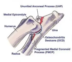
The "Big Three" Conditions
This mechanical overload leads to a cascade of joint failure, typically manifesting as one (or a combination) of three specific primary conditions inside the elbow:
1. Medial Coronoid Disease (FCP)
The most common manifestation. The chronic pounding on the overloaded medial coronoid process causes the bone beneath the cartilage to fatigue and develop microfractures. Eventually, the bone shatters, and fragments chip off inside the joint (Fragmented Coronoid Process). This acts like a sharp pebble trapped in a shoe, aggressively destroying the opposing cartilage.
2. Osteochondritis Dissecans (OCD)
A failure of the cartilage on the humerus to convert into normal bone during development. This leaves a thickened, weak flap of cartilage that can tear away from the bone, exposing sensitive nerve endings to the joint fluid.
3. Ununited Anconeal Process (UAP)
Common in breeds like the German Shepherd. The extreme pressure from an overgrown radius pushes up against the anconeal process (a peak of bone at the back of the ulna), preventing it from fusing to the main bone during adolescence. It remains a loose, unstable piece of bone inside the joint.
Diagnosis: Finding the "Footprints" on X-Rays
Because the canine elbow is so complex, standard 2D x-rays cannot always see the microscopic bone cracks or tiny chipped fragments floating inside the joint. The bones simply overlap too much on the image.
While CT scans are excellent at showing these tiny chips in 3D, we don't necessarily need a CT scan to know your dog needs help. When the elbow joint is suffering from chronic, uneven pressure, the bone reacts by thickening and hardening—a process called Subchondral Sclerosis.
When we look at your dog's standard x-rays, we are looking for this specific whitening and thickening of the bone. It acts as the "footprint" of the disease. If we see this sclerosis, combined with your dog's clinical lameness, it gives us all the diagnostic proof we need to proceed directly with an arthroscopic (keyhole camera) assessment to look inside the joint and treat the problem immediately.
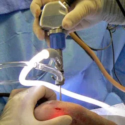
Treatment: Arthroscopy ("Keyhole" Surgery)
At BoneVet, we use advanced arthroscopy to treat elbow dysplasia. Instead of making a large, painful incision to open the entire joint, we insert a tiny, high-definition camera and micro-instruments through incisions no larger than a pen tip.
The Goals of Surgery:
- › Assess: We use the camera to magnify and inspect the joint cartilage in real-time.
- › Remove: We carefully extract any loose bone fragments causing immediate pain (removing the "pebble").
- › Resurface: We remove any cartilage flaps and cut or shave away the diseased, cracked bone of the medial coronoid process to eliminate ongoing "stress fractures" and pain, leaving a smooth surface that can form tough scar tissue.
Biomechanical Offloading
If a dog has significant joint incongruence, simply debriding the bone in the joint may not be enough. In some cases, we may perform an Ulnar Osteotomy (making a precise cut in the ulna bone). This allows the joint to dynamically shift and "settle" into a better, congruent fit as the dog heals, fundamentally altering the load distribution away from the damaged compartment.
The Honest Reality: Managing Lifelong Arthritis
Arthroscopy is highly effective at removing the immediate source of pain. However, we believe in being completely candid with our pet parents: surgery treats the damage, but it cannot fix the original genetic "poor fit" of your dog's bones.
Because of this, some degree of osteoarthritis will continue to develop over your dog's lifetime. Our goal with surgery is to dramatically improve comfort and function right now, and ideally slow the arthritic progression over the coming years.
A successful long-term outcome requires a partnership. Surgery is the vital first step, but it must be paired with your lifelong commitment to keep your dog lean, maintaining controlled exercise, targeted physiotherapy, and a tailored medical pain management strategy +/- high-grade joint supplements. Our goal is to give your dog the most comfortable, functional, and active life possible given the hand their genetics dealt them.